• A mosaic organism is composed of ≥2 genetically distinct cell populations derived from a homogeneous zygote.
– Genomic mosaicism results from alteration in the DNA sequence (affecting genes or chromosomes).
– Functional (epigenetic) mosaicism results from changes in gene expression (but not the DNA sequence) that are passed on during cellular replication; an important example is lyonization in female embryos, where random inactivation of one of the two X chromosomes occurs in each cell during early development.
• Clinical findings in mosaic skin conditions depend not only on the underlying genetic alteration but also on the timing of its origin (with earlier onset generally leading to more widespread involvement) and the cells or tissues affected (cutaneous ± extracutaneous).
– For example, mosaicism for an activating HRAS mutation can produce the combination of a nevus sebaceus (keratinocytes affected), speckled lentiginous nevus (melanocytes affected), and occasionally CNS abnormalities (nerve cells affected) in patients with phakomatosis pigmentokeratotica, a form of ‘twin spotting’.
• The accessibility of the skin allows visualization of mosaic patterns.
– Blaschko’s lines are streaks and swirls that represent pathways of epidermal cell (e.g. keratinocyte or melanocyte) migration during embryonic development (Fig. 51.1).
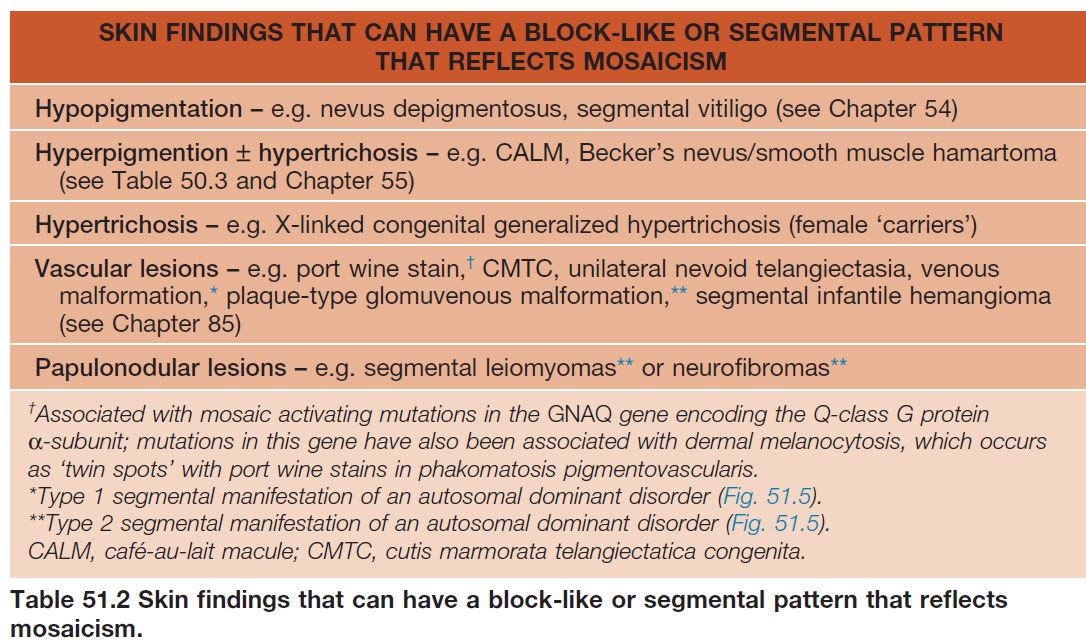
– Block-like, segmental, and dermatomal patterns can also reflect cutaneous mosaicism, typically involving melanocytes, mesodermal cells, and nerve cells, respectively.
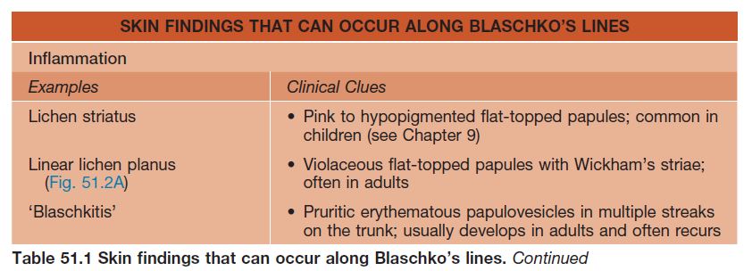
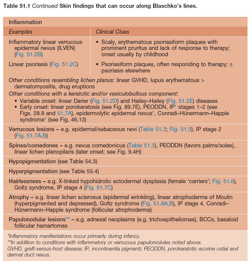
• Types of cutaneous lesions that can follow Blaschko’s lines or have a block-like/segmental pattern are outlined in Tables 51.1 and 51.2, respectively (Fig. 51.2).
• In chimerism, different cell populations reflect a genetically heterogeneous zygote (e.g. fusion of two zygotes or fertilization of one egg by two sperm); this may manifest with a block-like, linear, or irregular pattern of pigmentary variation.
Epidermal Nevi and ‘Epidermal Nevus Syndromes’
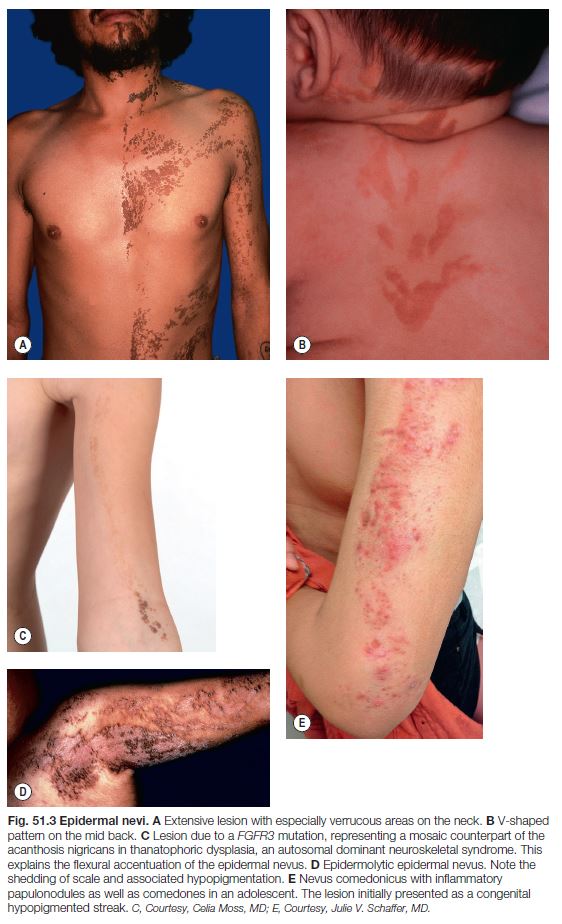
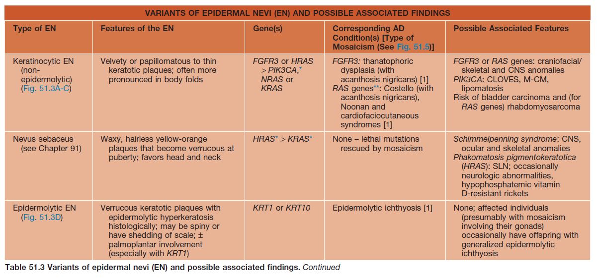
• Epidermal nevi (see Chapter 89) present as streaks and swirls of thickened (e.g. verrucous, hyperkeratotic, or velvety) skin along Blaschko’s lines, usually with hyperpigmentation and sometimes with adnexal involvement (e.g. in a nevus sebaceus or nevus comedonicus) (Fig. 51.3).
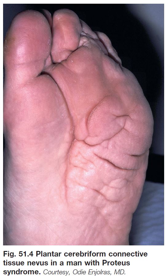
• The heterogeneous genetic etiologies and potential systemic associations (‘epidermal nevus syndromes’) of various types of epidermal nevi are presented in Table 51.3 (Fig. 51.4).
Mosaicism in Autosomal Dominant Skin Conditions
• When evaluating a patient with skin lesions in a mosaic pattern, it can be helpful to consider whether the findings would resemble an autosomal dominant genodermatosis if present in a more generalized distribution.
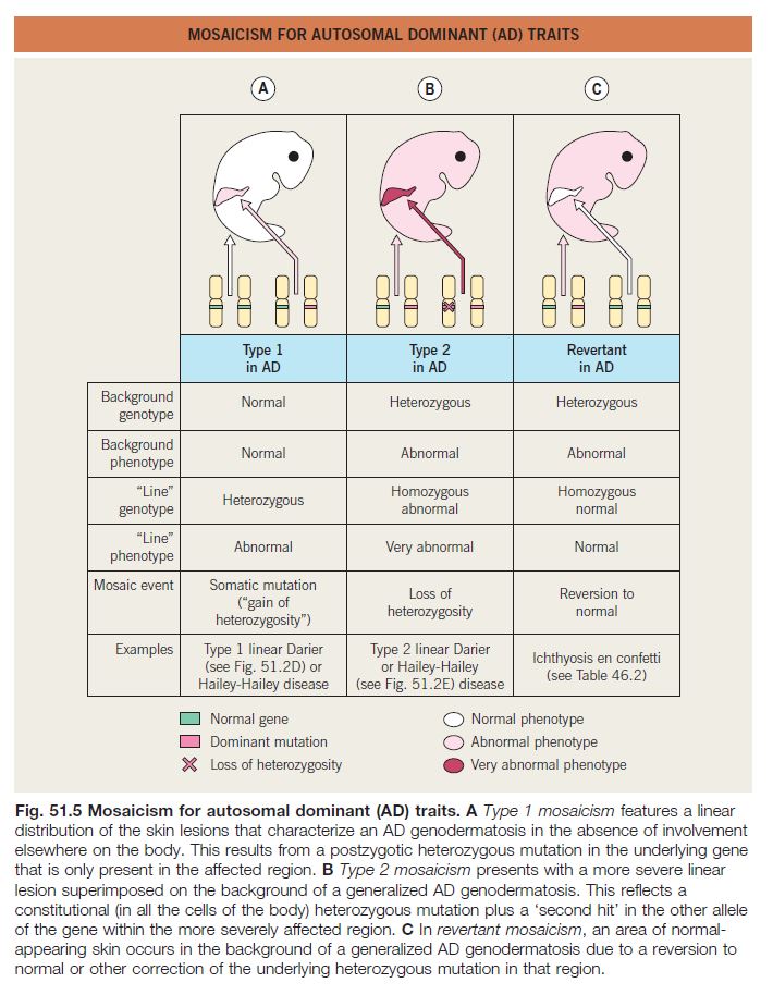
• Type 1, type 2, and revertant forms of mosaicism occurring in autosomal dominant skin disorders are summarized in Fig. 51.5.
Mosaicism in X-Linked Conditions
• In X-linked disorders, functional mosaicism due to lyonization occurs in female patients and carriers.
• X-linked dominant conditions are typically lethal in male embryos and are therefore seen almost exclusively in a mosaic pattern in female patients with a heterozygous mutation; male patients occasionally survive due to underlying mosaicism, e.g. in the setting of Klinefelter syndrome (47,XXY karyotype) or a postzygotic mutation.
• In conditions traditionally referred to as X-linked recessive, male patients have generalized disease and female patients or ‘carriers’ are affected to a variable (usually lesser) degree, often in a mosaic pattern; an example is hypohidrosis and hyperpigmentation following Blaschko’s lines in female ‘carriers’ of X-linked hypohidrotic ectodermal dysplasia (Fig. 51.6).
Incontinentia Pigmenti (IP)
• X-linked dominant disorder caused by mutations in the NF-κB essential modulator (NEMO) gene, which encodes a protein that protects against TNF-α-induced apoptosis; male patients with milder NEMO mutations may present with hypohidrotic ectodermal dysplasia with immune deficiency (see Table 52.5).
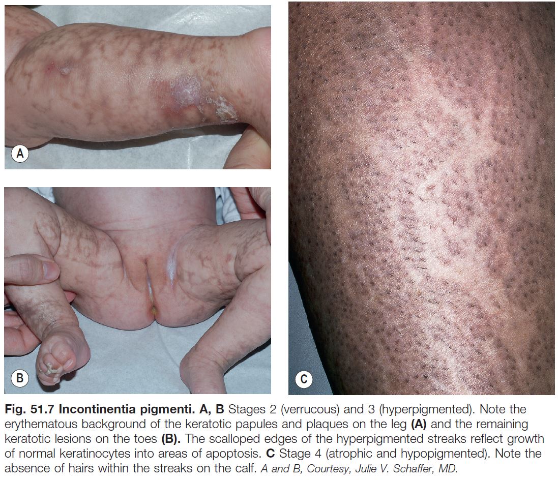
• IP has four stages, which may be absent or overlapping, of cutaneous findings that follow Blaschko’s lines (with the exception of stage 4) (Fig. 51.7; see Fig. 28.9):
– Vesicular streaks favoring the extremities and scalp in neonates, often associated with peripheral blood leukocytosis and eosinophilia; may recur during childhood febrile illnesses.
– Verrucous linear plaques favoring the extremities in infants; acral keratotic nodules sometimes develop after puberty.
– Hyperpigmented grayish-brown streaks and swirls favoring the trunk and intertriginous areas from infancy through adolescence.
– Hypopigmented/atrophic ‘Chinese character’-like bands lacking hair and sweat glands, favoring the calves in adolescents and adults.
• Other manifestations can include alopecia (often at the vertex in a swirled pattern), missing or conical teeth, retinal vascular anomalies (requires monitoring with ophthalmologic examinations during infancy), and CNS abnormalities (e.g. seizures, developmental delay).
Goltz Syndrome (Focal Dermal Hypoplasia)
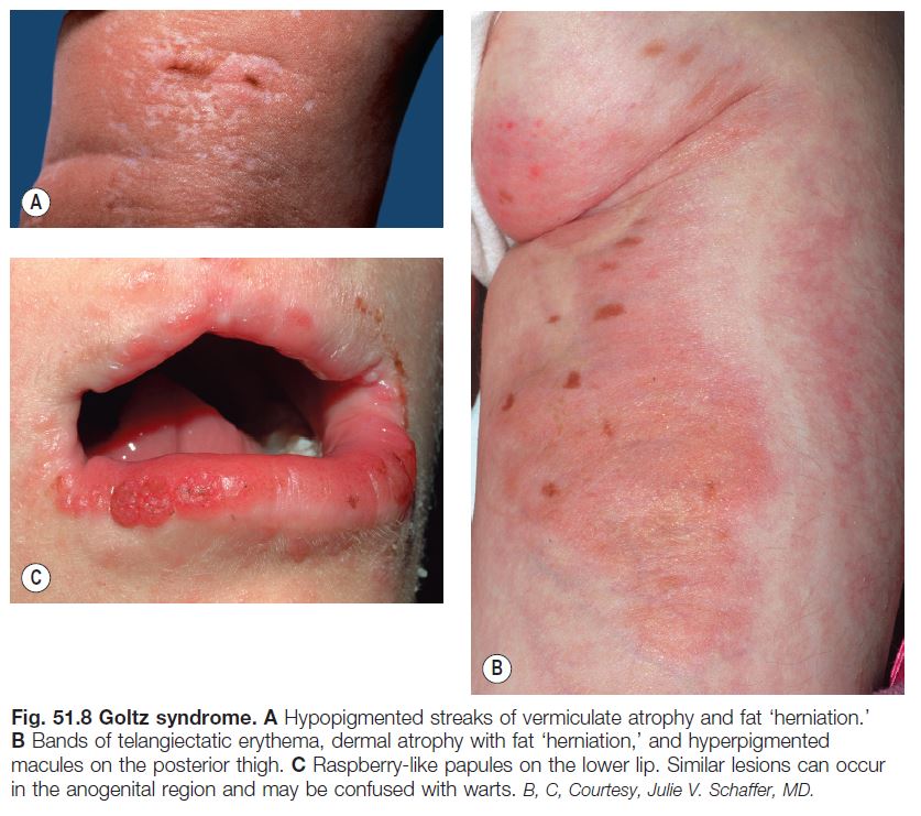
• X-linked dominant condition caused by mutations in the PORCN gene, which encodes a protein that regulates Wnt signaling.
• Skin lesions following Blaschko’s lines are characterized by vermiculate dermal atrophy, outpouchings of fat, telangiectasias, and hypo- > hyperpigmentation (Fig. 51.8).
• Additional features include periorificial raspberry-like papillomas, dystrophic nails, sparse hair, abnormal teeth, split hand/foot (‘lobster claw’) malformations, ocular abnormalities (e.g. microphthalmia), and the radiographic finding of osteopathia striata in long bones.
X-Linked Dominant Ichthyosiform Conditions
• Examples include congenital hemidysplasia with ichthyosiform nevus and limb defects (CHILD) and Conradi–Hünermann–Happle syndromes (see Table 46.2).
Lethal Disorders Rescued by Mosaicism
• Some mutations in autosomal genes are only observed in a mosaic state, as they would not be compatible with life if present in all the cells of the body.
• Examples include Proteus and Schimmelpenning syndromes presenting with epidermal and sebaceous nevi (see Table 51.3), Sturge–Weber syndrome (see Table 51.2 and Chapter 85), and forms of ‘pigmentary mosaicism’ including McCune–Albright syndrome (see Table 50.3 and Chapters 54 and 55).
Mosaic Manifestations of Acquired Skin Conditions
• Multifactorial skin disorders with environmental as well as genetic components occasionally occur along Blaschko’s lines or in a segmental distribution, presumably reflecting mosaicism for a ‘susceptibility’ mutation; as a result, such conditions often develop in older children or adults following an environmental trigger, and they are sometimes superimposed on a pre-existing mosaic lesion (e.g. psoriasis within an epidermal nevus).
• Examples include linear lichen planus, linear GVHD, and segmental vitiligo.

